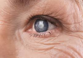
Refractive Lens Exchange (RLE) Replaces Your Eye’s Natural Lens With A Soft Gel Intra Ocular Lens (IOL) To Correct Your Refractive Error And Achieve Sharper Focus. Refractive Lens Exchange Is A Better Surgical Procedure Than Lasik Surgery For People Over 40 With Hyperopia.
Refractive Lens Exchange Surgery procedure is virtually identical to cataract surgery.
Patients over 40 with a deteriorating vision for distance (hyperopia) can benefit from refractive lens exchange to lessen their reliance on glasses. The difference between RLE and Cataract Surgery is that in refractive lens exchange, your lens being replaced is clearer, and your pre-surgery vision has not been reduced before surgery, by a cloudy cataract lens.
People who are middle-aged or older may have the beginnings of cataracts, that eventually could worsen, and require cataract surgery. Refractive lens exchange can be considered an excellent treatment for the early stages of cataract before a person suffers any vision loss due to a cloudy lens.
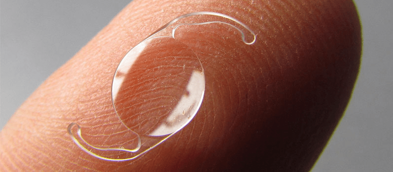
A new intra ocular lens is a permanent replacement for your natural lens and is designed to last the rest of your life.
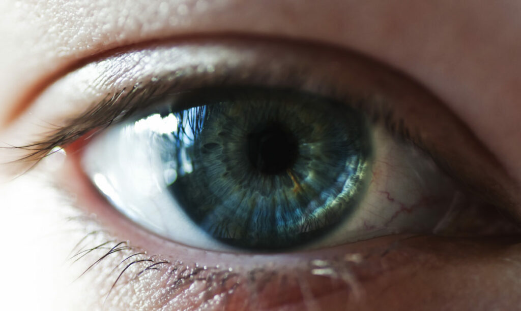
Since the lens is situated inside your eye and not on the surface like a contact lens, it is not visible to the naked eye, and you cannot feel it.
In the 1970/80’s RLE for cataracts was usually only performed if the patient was 20/70 vision or worse in the better eye. Now the guideline is “if a cataract is sufficient to interfere with the activities of daily living surgery is advised”
But, increasingly, people who have the initial stages of their lens starting to go cloudy ( with minimal vision loss) opt to do Refractive Lens Exchange surgery, while they are still physically healthy enough for surgery, even if their vision is not yet impeded by a cloudy lens. Refractive Lens Exchange Surgery is only done once per eye, so doing it early makes sense
Once a patient gets into their eighties other medical problems can sometimes make major surgery difficult .. and a person then has to put up with vision loss in later years, which could have been prevented by earlier RLE surgery.
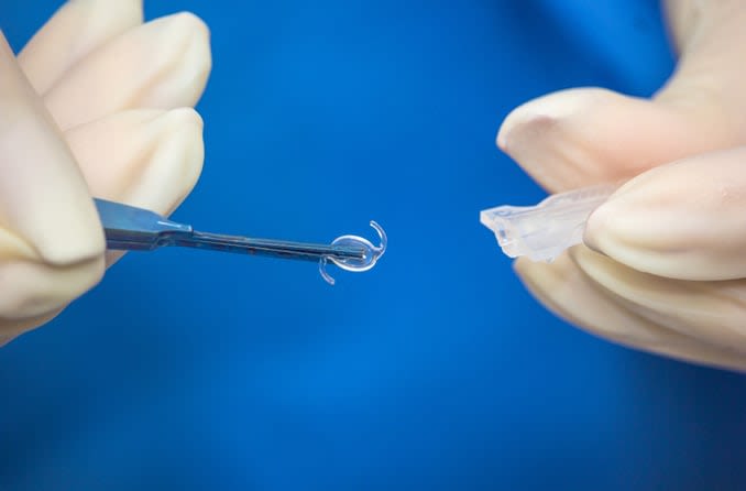
There are several choices of IOLs to insert during Surgery
Monofocal IOLs can be just distance and use glasses for reading. Or Multifocal IOLs can be one eye distance and one eye for reading (useful for people who have worn contact lenses in this way)
There are now different types of Multifocal IOLs. A multifocal lens that can be used for distance, intermediate, and reading. Or a newer form of multifocal IOL called an accommodating multifocal which works in the same way as the natural lens in your eye by contracting to see to read.
Recently given FDA approval as a MULTIFOCAL IOL, and achieving good results, is a new IOL called multifocal which has brought in a more advanced design in multifocal IOLS
Each multifocal IOL has advantages and disadvantages in terms of the best-uncorrected vision it produces at near, intermediate, and far distances, as well as the likelihood and degree of visual disturbances such as halos and night glare that might occur after surgery.
Should you as a patient go for, monofocal IOLs or multifocal IOLs?
There could be potential advances in technology which you may miss out on when you choose a new multifocal IOL today, “The future lens may offer improved quality of vision, but once you have refractive lens exchange that basically eliminates it as an option”
Or advances in technology may be many years away…it is difficult to make that judgment decision with a multifocal IOL, and will depend on various personal factors…. but if you are planning on having distance IOLs fitted and wearing reading glasses then the technology for these is excellent and an easy decision to make.
How do you find out if you are a good candidate for Refractive Lens Exchange?
The first step is to have an Eye Examination and then discuss with the Optometrist or the Ophthalmologist the possible options which are available to you for RLE, and if your age and your optical prescription are the most advisable for refractive lens exchange surgery.
You will be asked to do some general medical checks to confirm that you are OK for Surgery
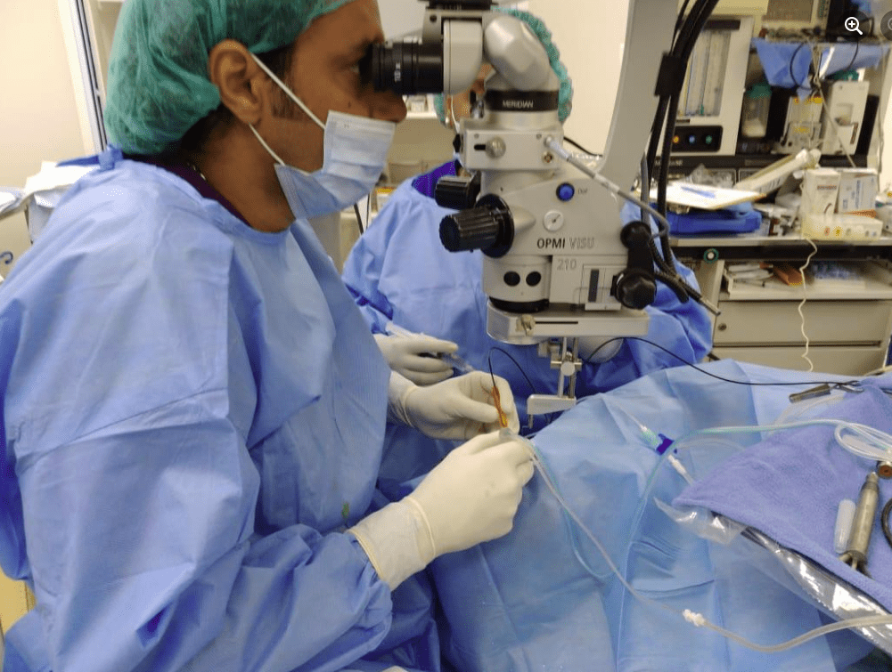
The VE Medical Surgery Centre offers a very modern, same-day, Private Surgery facility in Rodney Bay with a team with a lot of experience in RLE Surgery.
The Surgery team of Ophthalmologist, Surgery Nurse, Anesthetist, and Care Attendant provide excellent service to make your RLE experience as easy as possible.
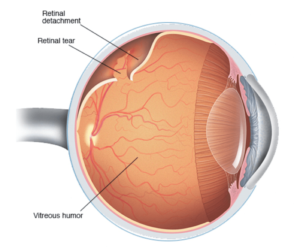
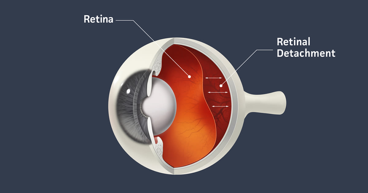
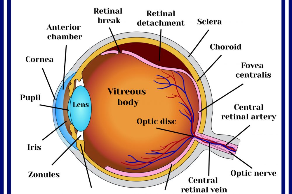
The retina is the light-sensitive tissue lining the back of the inside of the eye. It’s made up of different layers, including the light-sensing cells known as photoreceptors that are essential for sight. A layer of underlying supporting tissue contains blood vessels that supply the photoreceptors with the food and oxygen they need to stay healthy.
The retina functions as the line of communication between our eyes and the world around us by sending visual messages through the optic nerve to the brain. A healthy, intact retina is crucial for clear vision. But if it becomes detached, you can permanently lose your eyesight.
Types of Retinal Detachment
There are three ways a retina can detach:
Rhegmatogenous – A tear allows fluid to get under the retina, separating it from the retinal pigment epithelium (RPE), which nourishes the retina. This is the most common form of retinal detachment.
Tractional – Scar tissue on the retina’s surface contracts, causing it to separate from the RPE.
Exudative – Fluid leaks into the area underneath the retina. This type is usually caused by inflammatory diseases or eye injuries.
Can high blood pressure cause retinal detachment?
Along with causing heart and kidney problems, untreated high blood pressure can also affect your eyesight and lead to eye disease. Hypertension can cause damage to the blood vessels in the retina. This eye disease is known as hypertensive retinopathy.
What are the risk factors of a Detached Retina?
While retinal detachment is more common in people older than 40, especially men, it can occur at any age. Other risk factors include:
Family history
Having an eye disease or injury
Nearsightedness
Previous eye surgery
Weak areas in the retina
Symptoms of Retinal Detachment
Retinal detachment is painless; however, various warning signs point to its existence, including:
Blurred vision
Flashes of light in one or both eyes
Gradually reduced peripheral vision
Shadow over visual field
Sudden appearance of floaters
It is advised that should you experience any of these symptoms, you should seek medical attention immediately. At Vision Express, our ophthalmologist may diagnose retinal detachment via either of the following procedures:
Retinal Examination – In this procedure, a tool with a bright light and a special lens (ophthalmoscope) is used to examine the back of your eye, thus allowing your doctor to see retinal detachments, holes or tears.
Ultrasound Imaging – This procedure is used if bleeding has occurred in your eye making it difficult to see the retina.
Should your doctor fail to see a detachment, he or she may ask you to return in a few weeks to confirm that your eye has not developed any tears. Should you continue to experience symptoms, seek medical treatment immediately.
How is a Detached Retina treated?
Treatment for a detached or torn retina requires surgery. Tears are treated by sealing the retina to the back wall of the eye using these surgical treatments:
Freezing treatment (cryopexy): In this procedure, your eye surgeon uses a probe to freeze the retina around the tear. The scar helps secure the retina to the eye wall.
Laser surgery (photocoagulation): In this procedure, your ophthalmologist uses a laser to make small burns around the tear, creating scars. This seals the retina to the underlying tissue and helps prevent future detachment.
If your retina has detached, you will also need to have surgery to reattach it in its proper position. Otherwise, you could go blind. There are three surgical methods:
Pneumatic Retinopexy – In this procedure, a gas bubble is injected into the eye, pushing the tear back against the eye wall. Subsequently, your ophthalmologist will ask you to maintain a certain head position for several days.
Scleral buckle – In this procedure, a flexible band is placed around the eye to counteract the force pulling the retina out of place. Your doctor will drain the fluid underneath, allowing the retina to settle back into its normal position.
Vitrectomy – In this procedure, the vitreous gel is removed from the eye to prevent pulling. It is replaced with a gas or oil bubble to keep the retina in place until your body’s own fluids can fill the space. A gas bubble will go away on its own while an oil bubble must be surgically removed.
Following surgery, your vision may take months to improve and may not ever fully return. In some cases, the retina cannot be reattached and the eye will continue to lose sight.
How quickly should a Detached Retina be treated?
If your retina has detached, you will need surgery to repair it preferably within days of a diagnosis. The type of surgery your surgeon recommends will depend on several factors, including how severe the detachment is.
If you need to know more about retinal detachment, contact Vision Express today to make an appointment.
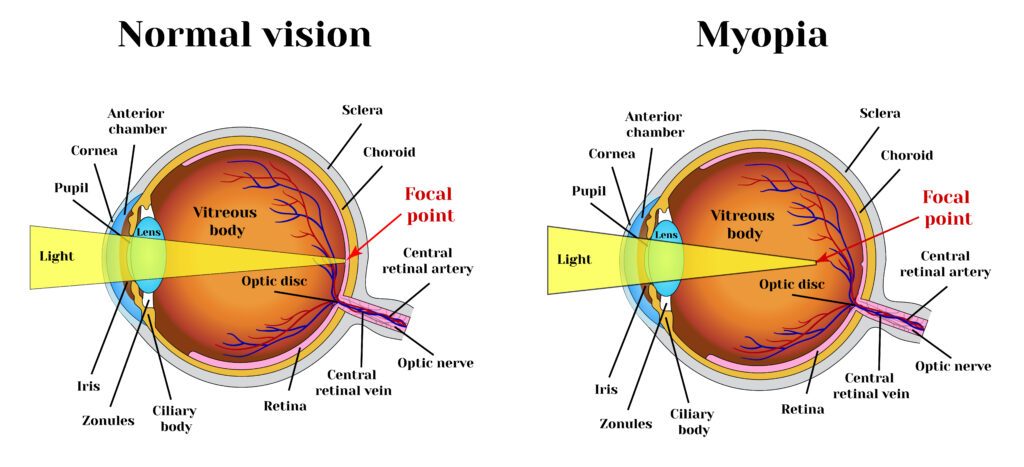
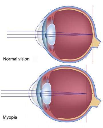
Myopia, also referred to as nearsightedness, is a vision condition whereby close objects can be seen clearly while distant objects appear blurred. As such, people with myopia have difficulty seeing a movie or the television screen clearly, a blackboard in class or while driving in traffic.
Myopia is caused when the eyeball is too long or the cornea (the clear front cover of the eye) is too curved, resulting in the light entering the eye to not focusing correctly and distant objects appearing blurred.
While the exact cause of myopia remains unknown, significant evidence suggests that many people inherit myopia or the tendency to develop the condition. There is an increased chance that children will be nearsighted if one or both parents are nearsighted.
While the tendency to develop myopia may be inherited, its actual development may be affected by how a person uses his or her eyes. People who spend considerable time reading, working at a computer, or doing other intense close visual work are more likely to develop myopia.
Myopia first occurs in school-age children. Since the eye continues to grow during childhood, it typically progresses until about age 20. However, myopia may also develop in adults due to visual stress or conditions such as diabetes.
Myopia can also be caused by environmental factors or other health problems:
Some people may experience blurred distance vision only at night. With “night myopia”, low light makes it difficult for the eyes to focus properly. Also, the increased pupil size during dark conditions allows more peripheral, unfocused light rays to enter the eye.
People who do excessive amounts of near-vision work may experience a false or “pseudo” myopia. Their blurred distance vision is caused by overuse of the eyes’ focusing mechanism. After long periods of near work, the eyes are unable to refocus to see clearly in the distance. Clear distance vision usually returns after resting the eyes. However, over time, constant visual stress may lead to permanent reduction in distance vision.
Symptoms of myopia may also be a sign of variations in blood sugar levels in people with diabetes or may be an early indication of a developing cataract.
At Vision Express, an optometrist can determine the cause of the vision problems through a comprehensive eye exam.
Diagnosis for myopia
Testing for myopia may use several procedures to measure how the eyes focus light and to determine the power of any optical lenses needed to correct the reduced vision.
As part of the testing, you will identify letters on a distance chart to measure visual acuity, written as a fraction, such as 20/40. The top number of the fraction is the standard distance at which testing is performed. The bottom number is the smallest letter size read. Someone with 20/40 visual acuity would have to get within 20 feet to identify a letter that could be seen clearly at 40 feet in a “normal” eye. Normal distance visual acuity is 20/20, although many people have 20/15 (better) vision.
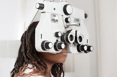
Using an instrument called a phoropter, the optometrist places a series of lenses in front of your eyes and measures how they focus light using a handheld lighted instrument called a retinoscope. The doctor may also use an automated instrument that evaluates the focusing power of the eye. The power is then refined based on your responses to determine the lenses that allow the clearest vision.
Your doctor can conduct this testing without using eye drops to determine how the eyes respond under normal seeing conditions. In some cases, such as for patients who can’t respond verbally or when some of the eye’s focusing power may be hidden, the doctor may use eye drops which temporarily keep the eyes from changing focus during testing.
Using the information from these tests, alongside the results of other tests of eye focusing and eye teaming, your doctor can determine if you have myopia and also the power of any lens correction needed to provide clear vision. Once testing is complete, your doctor can discuss treatment options.
Treatment for myopia
People with myopia have several options available to regain clear distance vision, including:
Eyeglasses: Eyeglasses are the primary choice for correction for most people with myopia. Depending on the amount of myopia, you may only need to wear glasses for certain activities such as watching a movie or driving a car. If you are very nearsighted, you may need to wear glasses all the time.
Generally, a single-vision lens is prescribed to provide clear vision at all distances. However, patients over age 40 or children and adults whose myopia is due to the stress of near-vision work may need a bifocal or progressive addition lens. These multifocal lenses provide different powers or strengths throughout the lens to allow for clear vision in the distance and up close.
Contact lenses: For some individuals, contact lenses offer clearer vision and a wider field of view than eyeglasses. However, since contact lenses are worn directly on the eyes, they require proper care to safeguard eye health.
Ortho-k or CRT: Another option for treating myopia is orthokeratology (ortho-k), also known as corneal refractive therapy (CRT). In this non-surgical procedure, you wear a series of specially-designed rigid contact lenses to gradually reshape the curvature of your cornea, the front outer surface of the eye.
The lenses place pressure on the cornea to flatten it and this changes how light entering the eye is focused. You wear the contact lenses for limited periods, such as overnight, before removing them. People with mild myopia may be able to temporarily obtain clear vision for most of their daily activities.
Laser procedures: Laser procedures such as LASIK (laser in situ keratomileusis) or PRK (photorefractive keratectomy) are also possible treatment options for myopia in adults. A laser beam of light reshapes the cornea by removing a small amount of eye tissue. The amount of myopia that PRK or LASIK can correct is limited by the amount of corneal tissue that can be safely removed.
In PRK, a laser removes a thin layer of tissue from the surface of the cornea in order to change its shape and refocus light entering the eye.
LASIK removes tissue from the inner layers, but not from the surface, of the cornea. To do this, a section of the outer corneal surface is lifted and folded back to expose the inner tissue. A laser then removes the precise amount of corneal tissue needed to reshape the eye. Then, the flap of outer tissue is placed back in position to heal.
Other refractive surgery procedures: People who are highly nearsighted or whose corneas are too thin for laser procedures may be able to have their myopia surgically corrected. A doctor may be able to implant small lenses with the desired optical correction in their eyes. The implant can be placed just in front of the natural lens (phakic intraocular lens implant), or the implant can replace the natural lens (clear lens extraction with intraocular lens implantation). This clear lens extraction procedure is similar to cataract surgery but occurs before a cataract is present.
Vision therapy for people with stress-related myopia: Vision therapy is an option for people whose blurred distance vision is caused by a spasm of the muscles that control eye focusing. Various eye exercises can improve poor eye focusing ability and regain clear distance vision.
If you have myopia, you have a variety of options to correct your vision problem. In consultation with your doctor at Vision Express, you can select the treatment that best meets your visual and lifestyle needs.
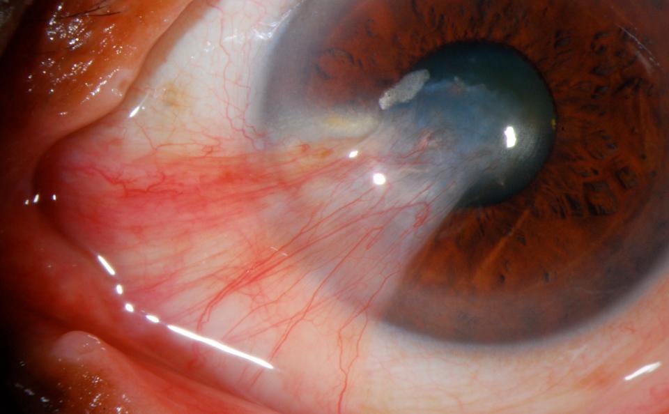
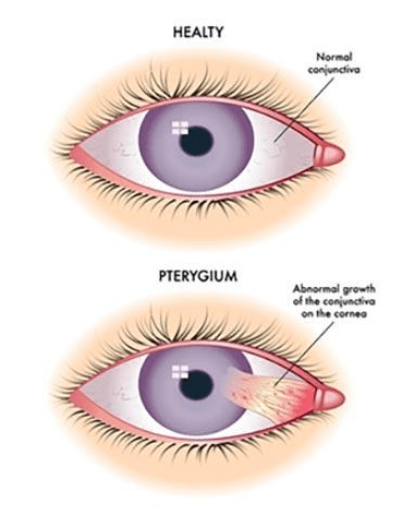
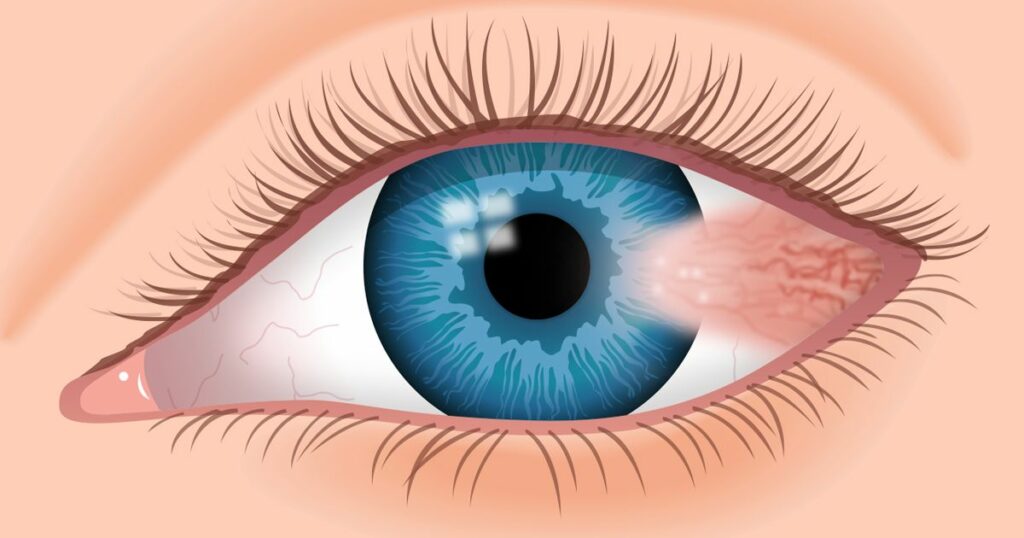
A pterygium is a growth of the conjunctiva or mucous membrane that covers the white part of your eye over the cornea. The cornea is the clear front covering of the eye. This benign or non-cancerous growth is often shaped like a wedge. A pterygium usually does not cause problems or require treatment, but it can be removed if it interferes with your vision.
While the exact cause of pterygium remains known, there is one theory that too much exposure to ultraviolet (UV) light can lead to these growths. The condition is more prevalent in people living in warm climates and spend a great deal of time outdoors in sunny or windy environments. Also, exposing your eyes regularly to certain elements increases your chances of developing pterygium. These elements include pollen, sand, smoke and wind.
While a pterygium does not always cause symptoms, when it does the symptoms are usually mild, including redness, blurred vision, and eye irritation. A burning sensation or itchiness might also occur.
A pterygium that grows large enough to cover your cornea can interfere with your vision. Moreover, a thick or larger pterygium can also cause you to feel like there’s a foreign object in your eye. Wearing contact lenses might be out of the question when you have a pterygium due to discomfort caused.
How serious is a pterygium?
Although a rare occurrence, a pterygium can lead to severe scarring on your cornea. Such scarring on the cornea will need to be treated because it can cause vision loss.
Treatment in minor cases usually involves eye drops or ointment to treat inflammation. However, in serious cases, treatment can involve surgical removal of the pterygium.
Diagnosing a pterygium is straightforward. At Vision Express, our eye doctor may diagnose this condition based on a physical examination using a slit lamp. This lamp allows the doctor to see your eye with the help of magnification and bright lighting. If your doctor needs to do additional tests, they may include:
Corneal topography: This medical mapping technique is used to measure curvature changes in your cornea.
Photo documentation: This procedure involves taking pictures to track the growth rate of the pterygium.
How is pterygium treated?
A pterygium does not usually require any treatment unless it’s blocking your vision or causing severe discomfort. At Vision Express, your eye doctor might want to check your eyes occasionally to see if the growth is causing vision problems.
What about medications?
If the pterygium is causing much irritation or redness, your doctor may prescribe eye drops or eye ointments that contain corticosteroids to reduce inflammation.
How about surgery?
Your doctor may recommend surgery to remove the pterygium if eye drops or ointments do not provide relief. Surgery is also done when a pterygium causes a loss of vision, or a condition called astigmatism, which can result in blurry vision. You can also discuss surgical procedures with your doctor at Vision Express if you want the pterygium removed for cosmetic reasons.
Obviously, there are risks associated with these operations. In some cases, a pterygium can return after being surgically removed. Your eye might also feel dry and irritated after surgery. Your doctor can prescribe medications to provide relief and reduce the risk of having a pterygium grow back.
As much as possible, avoid exposure to environmental factors that can cause a pterygium. Wear sunglasses or a hat to shield your eyes from sunlight, wind, and dust. Your sunglasses should also provide protection from the sun’s ultraviolet (UV) rays.
If you already have a pterygium, limiting your exposure to the following can slow its growth: wind, dust, pollen, smoke, and sunlight.
Avoiding the above conditions can also help prevent pterygiums from returning if you’ve had any removed.
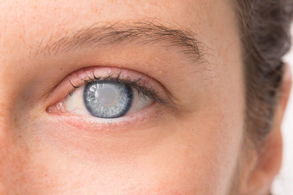
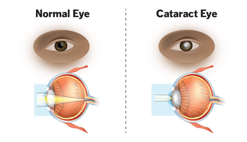
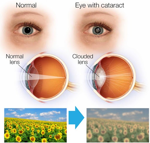
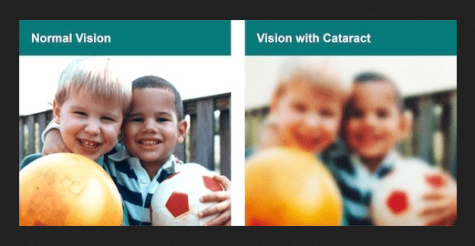
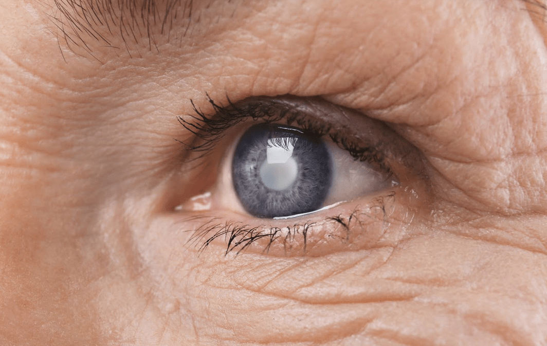
A cataract is a dense, cloudy area that forms in the lens of the eye. It begins when proteins in the eye form clumps that prevent the lens from sending clear images to the retina. The retina works by converting the light that comes through the lens into signals. The retina sends the signals to the optic nerve, which carries them to the brain.
While a cataract develops slowly over time, it
eventually interferes with your vision. Some people end up with cataracts in
both eyes; however, they usually don’t form simultaneously.
Cataracts are common in older people, with over 50%
of the people in the United States having cataracts or have undergone cataract
surgery by age 80.
Types of Cataracts
There are
different types of cataracts and they are classified based on where and how
they develop in your eye. They are:
Nuclear Cataracts: These form in the middle of the lens and cause the nucleus, or the centre, to become yellow or brown.
Cortical Cataracts: These are wedge-shaped and form around the edges of the nucleus.
Posterior Capsular Cataracts: These form faster than the previous two types and affect the back of the lens.
Congenital Cataracts: These, which are present at birth or form during a baby’s first year, are less common than age-related cataracts.
Secondary Cataracts: These are caused by disease or medications. Diseases that are linked with the development of cataracts include glaucoma and diabetes. The use of the steroid prednisone and other medications can sometimes lead to cataracts.
Traumatic Cataracts: These develop after an injury to the eye, but it can take several years for this to happen.
Radiation cataracts: These can form after a person undergoes radiation treatment for cancer.
What Causes Cataracts?
There are several underlying causes of cataracts, including:
An over-production of oxidants, which are oxygen molecules that have been chemically altered due to normal daily life
Smoking
Ultra-violet radiation
Long-term use of steroids and other medications
Certain diseases, such as diabetes
Trauma
Radiation therapy
Symptoms
of Cataracts
Common symptoms of cataracts include:
Blurry vision
Difficulty seeing at night
Seeing colours as faded
Increased sensitivity to glare
Halos surrounding lights
Double vision in the affected eye
The need for frequent changes in prescription glasses
Risk Factors of Cataracts
Risk factors associated with cataracts include:
Older age
Heavy alcohol use
Smoking
Obesity
High blood pressure
Previous eye injuries
Family history of cataracts
Over-exposure to the sun
Diabetes
Exposure to radiation from X-rays and cancer treatments
Diagnosing Cataracts
At Vision Express in Saint Lucia, where we perform
minor and major eye surgeries, your doctor will perform a comprehensive eye
exam to check for cataracts and to assess your vision.
Your doctor will put drops in your eyes to make
your pupils bigger, making it easier to check the optic nerve and retina at the
back of your eye for damage. Other tests your doctor might perform include
checking your sensitivity to glare and your perception of colours.
Prevention of Cataracts
To reduce your risk of developing cataracts, please
follow these crucial steps:
Protect your eyes from UVB rays by wearing sunglasses outside
Have regular eye exams
Quit smoking
Eat fruits and vegetables that contain antioxidants
Maintain a healthy weight
Keep diabetes and other medical conditions in check
Treatment of Cataracts
If you’re unable or uninterested in surgery, Vision
Express will help you manage your symptoms. However, our very attractive
payment plans ensure that you get the right care you need. We can also suggest
stronger eyeglasses, magnifying lenses, or sunglasses with an anti-glare
coating.
Surgery
Surgery is recommended when cataracts prevent you
from going about your daily activities, such as reading or driving. It’s also
performed when cataracts interfere with the treatment of other eye problems.
Extra-capsular surgery involves removing the cloudy
part of the lens through an incision in the cornea. After surgery, an
artificial intraocular lens is placed where the natural lens was.
Cataracts can interfere with daily activities and
lead to blindness when left untreated. Cataract surgery is one of the most
successful procedures in the world.
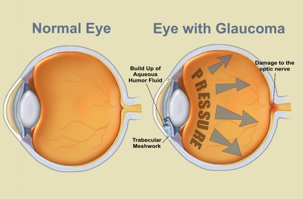
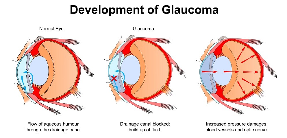
Like a thief in the night,glaucomacan rob you of your vision, slowly damaging your eyes and causing irreparable harm. Despite having been around for eons, researchers remain puzzled by what causes glaucoma in most cases. While available treatments can delay vision loss, there is no cure for glaucoma, resulting in the eye condition being a leading cause of blindness globally.
By definition, glaucoma is a group of diseases that damage
the optic nerve, a cable at the back of each eye that connects it to the brain.
It affects more than 60 million worldwide. While there are various forms of the
disease, primary open-angle glaucoma is the most common and most mysterious.
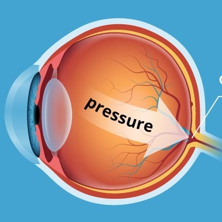
Anyone who has had an air-puff test — otherwise known as tonometry – may also know that glaucoma is linked to an increase in eye pressure, known as intraocular pressure. The eye’s unique anatomy, in combination with other factors, can increase eye pressure, leading to some types of glaucoma.
The front part of the eye, between the cornea — the eye’s front window — and the iris — the colored part of the eye — is filled with a clear fluid. This fluid leaves the eye, entering the blood via a gap at the angle where the cornea and iris meet. This gap is filled with a sponge-like tissue known as the trabecular meshwork which helps to regulate fluid passage. Often, eye infections, injuries or certain medications narrow the gap and compress this spongy tissue, producing a rapid build-up of fluid and eye pressure. This is referred to as angle-closure glaucoma.
Open-angle glaucoma is more common than angle-closure glaucoma. It has a more gradual course and there are no clear signs of blockage within the eye’s drainage system. However, researchers estimate that between 50% and 80% of people with open-angle glaucoma have eye pressure that is higher than average. Others have normal pressure or even low pressure. Conversely, many people have high eye pressure, yet never develop glaucoma.
Elevated intraocular pressure is a leading risk factor for primary open-angle glaucoma. The higher the intraocular pressure, the more likely the person is to develop glaucoma and the more likely it is to progress.
Medications that lower eye pressure are vital for glaucoma treatment. In many instances, a doctor would recommend surgery to increase fluid drainage from the eye. Drugs, surgery, or both approaches together are often successful at slowing the course of open-angle glaucoma. Vision Express has several treatments options.
Pressure-lowering eye drops have also been found to delay the onset of glaucoma in people with high eye pressure. But even with medication or surgery, open-angle glaucoma usually continues to attack the optic nerve and cause gradual vision loss.Vision Express has state of the art equipment to test and evaluate your eye health.
Besides high eye pressure, other risk factors for open-angle glaucoma exist and might provide clues about what causes it. Age, for example, is a clear risk factor. Another example: open-angle glaucoma is rare among Americans under age 50, but affects nearly 8% of Americans over 80.
Long before we grow old and even before we’re born, other
factors come into play, including ancestry. Open-angle glaucoma is about five
times more common among African-Americans and Mexican-Americans compared to
Whites, and has an earlier, more rapid course in African- Americans. Family
history also has a strong influence. A person’s risk of developing open-angle
glaucoma is about 10 times higher if a parent or sibling has it, and the risk
is higher still if an identical twin has it.
As researchers continue to dig deeper into the genetics of
open-angle glaucoma, sometimes open-angle glaucoma is passed from parent to
child due to defects within a single gene. Eight such genes have been
identified so far, and they tend to cause glaucoma with an early onset before
age 50. Due to these discoveries, there are now genetic tests that can help
people with early-onset glaucoma determine the risk that their children will
inherit the disease.
Generally, studies have shown that people are living longer these days and there seems to be a direct correlation between a healthy lifestyle and minimal years of disability. In those studies, it was determined that people with healthy lifestyles lived longer and also experienced fewer years of being ill compared to those not adopting healthy lifestyles. This should be enough reason to inspire people to adopt a healthy lifestyle because it’s never too late to start doing something good.
There are many ways to improve one’s health through a healthy
diet and exercise — even well into old age. In fact, lack of physical activity
and regular exercise coupled with poor diet can contribute to high blood
pressure, heart disease and heart attacks, lack of sexual activity, and a poor
health-related quality of life.
In order to truly appreciate the value of a healthy lifestyle as
older adults, it’s important to acknowledge the physical and emotional changes
that occur within us as we age. Having such awareness of the changes reduces
the surprise effect. On the emotional level, we struggle with the loss of loved
ones, careers, and independence. On the physical level, our bodies are less
vibrant as they were when we were younger.
These changes notwithstanding, we can still live a quality and
fulfilling life. Below are some important ways to live your life during your
older years by maintaining a healthy lifestyle.
Diet Is Very Important
It is a very important for seniors to practice healthy eating habits. But as you advance in age, some noticeable changes will occur, including slower metabolism and changes in your sense of smell and taste which can ultimately affect your appetite.
Shopping for appropriate food and preparing it will not be a
walk in the park, either. If this occurs, it’s advisable to reach out to family
members or your health provider for assistance. There are also community programmes
that provide healthy food to seniors.
If you are able to prepare your own food, it’s important that
your diet is rich in fibre, vegetables, fruits, whole grains, and lean protein
(meat) which keep you energetic and aid your slow digestion.
Making your food taste and look good also encourages your
appetite. Always stay hydrated — even when you do not feel thirsty — as water
keeps your energy levels up and makes your skin smooth and looking young. If
possible, eat with friends, family, or neighbours; this allows you to keep in
touch with them and be more encouraged to eat despite your reduced appetite.
Eating right also helps you to maintain a healthy weight, which decreases
your risk of certain types of arthritis and diabetes. Avoid smoking cigarettes since
they are seriously harmful to your body and predispose you to lung cancer,
heart disease, osteoporosis, and bronchitis. The chemicals in cigarettes also
damage the skin, causing you to look older than you really are.
Stay Active!
Studies have found that exercise is a top contributor to living longer as it adds more years to your life. If you have not been exercising before, don’t be too hard on yourself because it is never too late to start. Just start!
The benefits of exercise are numerous and rewarding:
Prevents memory loss (dementia)
Produces eel-good hormones (endorphins)
Helps to reduce chronic pain
Increases muscle mass from weight training and improves metabolism
Improves quality of sleep
Improves flexibility, balance, and good posture
Boosts the immune system
Practicing
yoga also relieves discomfort caused by arthritis and fibromyalgia. So before
you enroll in any exercise programme, be sure to consult your healthcare
provider. Start out small and build up slowly over time. Do something you enjoy
to keep you motivated, such as cycling, golfing, walking a pet, gardening, or
swimming.
Exercise
with a friend or a family member or join a class; that way, you keep each other
motivated. Yoga is also a great form of exercise. Muscles stiffen and shorten
when we do not exercise, but stretching improves that. So try stretching for at
least five minutes daily and you will notice the difference.
Exercise The Brain!
Keep your brain active by feeding your creativity, especially now that you are retired and no longer challenged by your career. While physical exercise helps keep your brain alert, other activities like word games, crossword puzzles, learning a new language, and learning a new skill can keep your brain strong.
Stay Positive And
Stay Connected
There
are many difficult challenges that come with getting older, such as losing
loved ones, your independence, and your health. Despite all these things, you
must remain strong and try to navigate through these challenges. Below are some
few tips you can follow:
Accept the thingsthat have changed in your life, even though
this will be difficult. However, accepting and letting go of things out of your
control reduces your stress levels.
Remind yourself of the things
that you are still grateful for, such as the family and friends.
Reach out to other people who
may be experiencing similar issues as yours. Join a support group or volunteer
in the community to help you focus less on your own hardships. Socialize with
people and spend time with at least one person daily. Physical interactions
ward off depression and are much better than texts or emails. Make new friends.
Although your older friend may have moved away or died, you can still make new
ones and share memories with them.
Acknowledge how you feel, especially
after the loss. Don’t bury your emotions; facing them is the only healthy way
to work through them. Talk to a friend, family member, or professional. Keeping
a journal can also be helpful.
Maintain a sense of purpose.
Retirement does not mean your life is without purpose. You still have a lot to
contribute to the world. Now is a great time to write your memoirs, reflect on
your life experiences and the lessons you learned from them. You now have more
time to spend time with your family members. Take up a new hobby or focus on an
old one. Travel and see your city or your country or learn a new language,
sport or musical instrument. Visit the museum, go to a concert, or a play.
These activities will help you maintain your sense of joy.
Sleep comfortably. Many adults suffer
from sleep problems as they get older, including waking up often during the
night, insomnia, and daytime sleepiness. Low quality sleep is caused by poor
sleeping habits, but you can use the following tips to improve that:
Keep Artificial Light To A
Minimum. Artificial light suppresses
the production of hormones responsible for causing sleepiness called melatonin.
Switch off the television and computer an hour before you sleep and use
low-watt bulbs.
Improve Bedtime Rituals. Take a warm relaxing bath or listen to slow music before your
bedtime.
Create A Space That Is
Conducive To Sleeping. Invest in a comfortable
mattress and ensure your bedroom is dark, quiet and cool.
Exercise Regularly. Especially a few hours before bedtime promotes better sleep.
Prevention Is
Better Than Cure
There are several precautionary measures you can take to improve the quality of your life, including:
Seeing your health professional on a regular basis and following their recommendations on screening and preventive measures. These may include yearly flu shot, screening for breast cancer, cervical cancer, checking blood pressure, etc.
Paying
attention to your body and alerting your healthcare
provider immediately if something feels off. For instance, if you feel dizzy or
unsteady, it’s important to follow up on this with your doctor to avoid a fall.
According to the Center for Disease Control and Prevention (CDC), falls were
listed as the first cause of death-related injury among seniors.
Keeping
your home safe by making sure that all rooms
are well lit, moving furniture that can be an obstruction, ensuring that electrical
and gas appliances are safe and up-to-date, looking out for wiring that’s loose
or rugs or carpets that can cause a fall. Also ensure that your home is
properly insulated.
At Vision Express,
we do our best to ensure that we not only give you the information you need as
it relates to your eyes, but our expert and dedicated team also goes above and
beyond to ensure that you see the world clearly. Part of our job is to also
provide answers to the many questions you have about your eyes.
Question: I have been told I have a cataract. What happens now?
Answer: If you have been told that you have a cataract, there is no need to be overly
concerned as you are not alone. Each year, millions of people worldwide have
cataract surgery. Thanks to advanced surgical procedures and technology,
cataract surgery is not only one of the most frequently performed surgical
procedures, but also one of the safest and most successful surgical procedures
that you can have. At Visio Express, cataract surgery is performed on an out-patient
basis and usually only requires a few hours of your time from beginning to end.
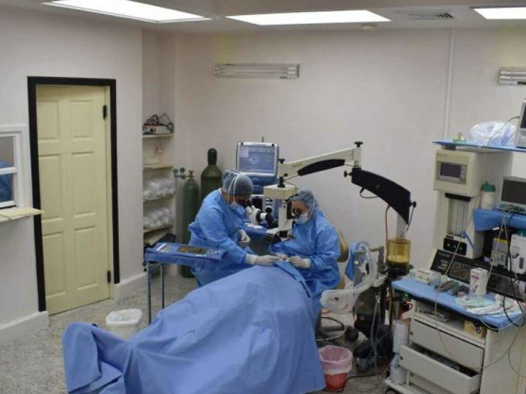
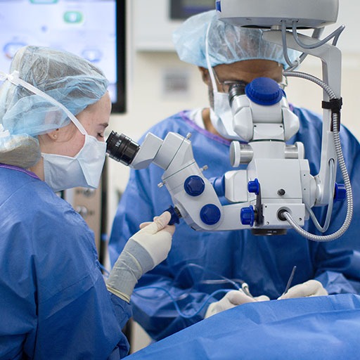
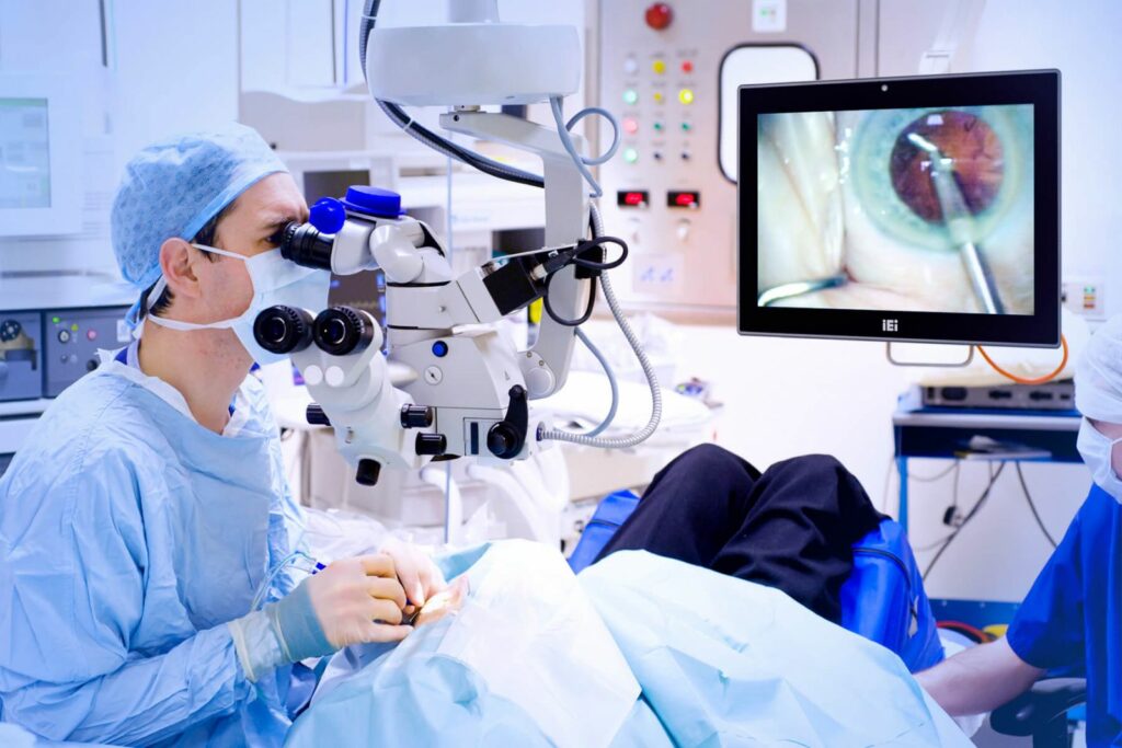
Cataract procedure can be performed under local anesthetic. At Vision Express Surgery Centre, the team on the day of surgery comprises the Ophthalmologist, the Ophthalmic Surgery Nurse, the Anaesthetist and the Pre- and Post-Surgery Caregiver.
Question: What happens on Surgery Day?
Answer: When you arrive at VE Medical Centre in Rodney Bay, and change into a surgery
gown, the nurse will begin by placing eye drops in your eyes to dilate your
pupil and numb and anesthetize the surface of your eye. Several sets of eye
drops may be administered for Cataract Surgery. The use of eye drop anesthesia
is sufficient for most cataract patients so that they feel just about nothing
and experience little, if any, discomfort at all.
During your Cataract Surgery, you will be
aware of the surgeon, the staff and the operating room surroundings, but will
not be able to see images or the surgery being performed. Expect to see team
members in scrub uniforms and surgical gowns, and wearing masks to protect the
sterile area. During the surgery, you should not experience pain and minimal,
if any, discomfort.
The procedure involves a very small cut
being made in the eye, the cloudy cataract lens is removed and the new IOL
inserted. The VE Medical team also use the phaco-emulsifaction equipment to
remove the lens and clean out the lens cavity. The cut to remove the lens made
by the Surgery Team at Vision Express is very small and normally requires no
stitches. Your surgeon will complete the surgery by placing some antibiotic
drops and possibly some anti-inflammatory drops in your eye to prevent
infection and swelling.
The surgery takes less than an hour. After
the surgery, you will rest for a while and then go home. The surgeon will
normally ask you to wear an eye patch for a couple of days and you will need to
use eye drops to reduce any possibility of infection. You will return for a
post-surgery visit 24 hours after the surgery and then a week later for a final
post-surgery check-up.
Here’s a review from Linda Hastilow,
following her Cataract Surgery performed in January 2016 at VE Medical: “I had a cataract operation yesterday, and
must praise the surgeon Dr. Alejandro and the staff. Their attention to detail
with care and comfort was superb. The impressive part is the operating room, so
well-equipped with hi-tech equipment and highly-trained staff. From what was a
very nervous patient, not anymore — now full of confidence.”
Question: Which Intraocular lens is right for me?
Answer: There are several choices of IOLs to insert during Surgery — Monofocal IOLs
can be just distance, and you use glasses for reading. This is the choice most
cataract patients make. They are comfortable to have back clear distance vision
and are happy to have a pair of glasses for reading or there are now different
types of Multifocal IOLs. A multifocal lens can be used for distance,
intermediate and reading. Or a newer form of multifocal IOL called an
accommodating multifocal which works in the same way as the natural lens in
your eye by contracting to see to read.
Recently given FDA approval as a
multifocal IOL and achieving good results is a new IOL called Technis Symfony,
which has brought in a more advanced design in multifocal IOLS. Each multifocal
IOL has advantages and disadvantages in terms of the best uncorrected vision it
produces at near, intermediate and far distances, as well as the likelihood and
degree of vision disturbances such as halos and night glare that might occur
after surgery.
When you come for your pre-cataract
surgery appointment, your Ophthalmologist at Vision Express will discuss the
options and make the best suggestion. Full measurements will be taken to get
the correct IOL for you.
Question: I think my vision is getting cloudy from a
cataract. What do I do?
Answer: Step one is to go for a full eye examination with the Optometrist who will tell
you the cause of your cloudy vision. If it is a cataract, then relief is at
hand because cataract surgery can restore your vision. The Ophthalmologist will
do a full pre-cataract surgery examination and you will need to go for some
routine medical checkups to confirm you are okay for surgery. Then just plan
the surgery day and let the excellent surgery team at VE Medical in Karlione
Court, Rodney Bay, take care of the rest.
Frequently Asked
Questions (FAQs) About Cataract Surgery and Pre-Surgery
Question: What will happen at the pre-surgery?
Answer: At your appointment with Dr. Garcia, we will do a full assessment of your eye
health and complete a lens calculation. We will also check your lens
prescription and order this accordingly. The date of your procedure will also
be confirmed.
Question: Will my GP require any
forms to be signed?
Answer: Yes. At the
pre-surgery appointment, we will give you forms for your GP to complete in
order to confirm your general health is well enough for surgery.
Question: Will
you check my blood pressure at the pre-appointment?
Answer: Certainly. This is a vital part of your health screening. We will also give you an overview of ‘do’s and ‘don’ts’ so you are in the best possible health for your surgery.
Question: What time will my surgery take place?
Answer: We only perform day surgery. Your appointment will either be in the morning or
the afternoon.
Question: Will
I need someone to take me home?
Answer: Yes. You will not be able drive and highly recommend you have a friend or loved one to assist.
Question: What about my partner/friend? Will they need to wait throughout the surgery?
Answer: No. We will call and text them half an hour before the surgery finishes or
whenever is convenient to them.
Question: Will
I need to come back?
Answer: Yes. We have a post-surgery appointment to check your progress and to confirm your eye health is in excellent condition.
Answer: Yes. The price is inclusive of the pre-surgery, surgeon fees, lens implant and the post-surgery appointment.
Question: How much deposit do I need to put down?
Answer: We normally ask for a 25%
deposit at the pre-surgery appointment and the balance on the day of the
appointment if paying in full. Discuss your payment options with the team at
Vision Express.
BUT Cataract Surgery is covered by medical
insurance. If you don’t have medical insurance coverage, then Vision Express
can arrange an interest-free payment plan with your Credit Union, or a two-year
Vision Express “Vision Plan” with FastCash.
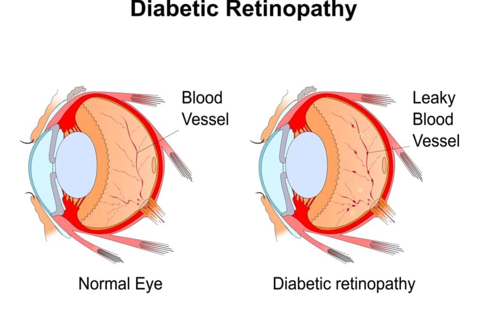
An eye
condition that can cause vision loss and blindness in people
with diabetes, diabetic retinopathy affects
blood vessels in the retina, the light-sensitive layer of tissue in the
back of the eye.
It’s important for anyone diagnosed with diabetes to get a comprehensive dilated eye exam at least once a year. While diabetic retinopathymay not present any symptoms initially, in later stages of the disease blood vessels in the retina start to bleed into the vitreous (gel-like fluid in the centre of the eye).
If
this happens, you may see dark, floating
spots or streaks that look like cobwebs. Sometimes, the spots
clear up on their own, but it’s important to get treatment
immediately. Without treatment, the bleeding can recur, get
worse, or cause scarring. Therefore, early detection is crucial in
determining the steps you need to take to protect your vision.
Diabetic retinopathy is caused by high blood sugar due to diabetes. Over time, having too much sugar in your blood can damage your retina, the part of the eye that detects light and sends signals to the brain through a nerve in the back of the eye (optic nerve).
Diabetes
damages blood vessels throughout the body. The damage to the
eyes starts when sugar blocks the tiny blood vessels that go
to the retina, causing them to leak fluid or bleed. To make
up for these blocked blood vessels, your eyes then grow new blood vessels
that do not work well. These new blood vessels can leak or bleed easily.
Complications
Diabetic retinopathy involves the abnormal growth of blood vessels
in the retina. Complications can lead to serious vision problems, including:
Vitreous hemorrhage: The new blood vessels may bleed into the clear, jelly-like substance that fills the centre of your eye. If the amount of bleeding is small, you might see only a few dark spots (floaters). In more severe cases, blood can fill the vitreous cavity and completely block your vision. Vitreous hemorrhage by itself usually doesn’t cause permanent vision loss. The blood often clears from the eye within a few weeks or months. Unless your retina is damaged, your vision may return to its previous clarity.
Retinal detachment: The abnormal blood vessels associated with diabetic retinopathy stimulate the growth of scar tissue, which can pull the retina away from the back of the eye. This may cause spots floating in your vision, flashes of light or severe vision loss.
Glaucoma: New blood vessels may grow in the front part of your eye and interfere with the normal flow of fluid out of the eye, causing pressure in the eye to build up (glaucoma). This pressure can damage the nerve that carries images from your eye to your brain (optic nerve).
Blindness: Eventually, diabetic retinopathy, glaucoma or both can lead to complete vision loss.
It’s worth noting that while diabetic retinopathy is the most common cause of vision loss for people with diabetes, diabetics also run the risk of developing other eye conditions, including cataracts, open-angle glaucoma, diabetic macular edema, neovascular glaucoma, and retinal detachment.
A cataract is a clouding of the lens of the
eye which leads to decreased vision. Cataracts often develop
slowly and can affect one or both eyes. Symptoms may include faded colours,
blurry or double vision, halos around light, trouble with bright lights, and
trouble seeing at night. Having diabetes makes
you 2 to 5 times more likely to develop cataracts. It also
makes you more likely to get them at
a younger age.
In open–angle glaucoma, the angle in your eye where the iris
meets the cornea is as wide and open as
it should be. However, the eye’s drainage canals become clogged over time,
causing an increased internal eye pressure and subsequent damage to the optic
nerve. Having diabetes nearly
doubles your risk of developing a type of glaucoma called
open-angle glaucoma.
Over time, nearly 50% of people with diabetic retinopathy will
develop diabetic macular enema,
or DME, which occurs when blood
vessels in the retina leak fluid, causing swelling in the macula
(a part of the retina). People with DME will experience blurry vision
because of the extra fluid in their macula.
Diabetic retinopathy can
cause abnormal blood vessels to grow out of the retina and block
fluid from draining out of the eye. This causes a type of glaucoma, neovascular
glaucoma.
Diabetic retinopathy can cause scars to form in the back of your eye. When the
scars pull your retina away from the back of your eye, it’s
called tractional retinal detachment.
Prevention
Managing diabetes is
the best way to lower your risk of diabetic
retinopathy. This means keeping your blood sugar levels as close
to normal as possible. You can do this by getting regular physical activity, eating healthy
and carefully following your doctor’s instructions for your insulin
or other diabetes medication.
To help control
your blood sugar, you’ll need a special test called an A1c test,
which shows your average blood sugar level over a three-month
period. Consult your doctor about lowering
your A1c level to help prevent or manage diabetic retinopathy.
Having
high blood pressure or high cholesterol along with diabetes increases
your risk for diabetic retinopathy. By
controlling your blood pressure and cholesterol, you’re also helping to
lower your risk for vision loss.
The
diagnosis for diabetic retinopathy is
having a dilated eye exam. Treatment include medication, laser treatment, and
surgery. Staying physically active, eating healthy
and taking your medication are key ways to manage your diabetes and
help prevent or delay vision loss.
Injections: Medicines called anti-VEGF drugs can slow
down or reverse diabetic retinopathy. Other medicines,
called corticosteroids, can also help.
Laser treatment: To reduce swelling in
your retina, eye doctors can use lasers to make the
blood vessels shrink and stop leaking.
Eye surgery: If your retina is bleeding a lot or you have a
lot of scars in your eye, your eye doctor may recommend a
type of surgery called a vitrectomy.
Patients with diabetes who successfully manage their blood sugar
levels will help to prevent the onset of a severe form of diabetic retinopathy. They can achieve this by following these
simple rules:
Eating a healthy and
balanced diet
Exercising regularly
Maintaining a healthy
body weight
Quitting smoking
Strictly controlling
alcohol intake
Taking any anti-hypertensive
measures according to their doctor’s instruction
Having regular eye
examinations to check and monitor any problems early
Who’s
at risk of developing diabetic retinopathy?
Anyone
with any kind of diabetes can get diabetic
retinopathy, including Type I, Type II and gestational diabetes
(diabetes that can develop during pregnancy). Your risk increases the
longer you have diabetes.
More than 2 in every 5 Americans with diabetes have some stage of diabetic retinopathy. Fortunately, you can lower your risk of developing diabetic retinopathy by controlling your diabetes.
Women with diabetes who become pregnant — or women who develop gestational diabetes — are at high risk for getting diabetic retinopathy. Therefore, women with diabetes and are pregnant should have a comprehensive dilated eye exam as soon as possible. They should also ask their doctor as to whether they will need additional eye exams during their pregnancy.
At
Vision Express, our Retinal Ophthalmologist,
Dr. Alejandro Garcia, checks for diabetic
retinopathy as part of a dilated eye exam. The exam
is simple and painless. The doctor will give you some eye drops to dilate
(or widen) your pupil and then check your eyes for diabetic retinopathy and other eye
problems.
Vision Express has a
regular diabetic clinic at the Rodney Branch to help with checking your blood
sugar and advice on your diet. If you are diagnosed with diabetes, an annual
eye examination is very important. With diabetic
retinopathy, vision once lost cannot be restored, but further vision loss
can normally be prevented.
The retina in your eye
has tiny blood vessels that are easy to damage. Having high blood glucose and
high blood pressure for a long time can damage these tiny blood vessels. Diabetic
retinopathy affects up to 80% of those who have had diabetes for 20
years or more. At least 90% of new cases can be reduced with proper treatment
and monitoring of the eyes. The longer a person has diabetes, the higher his or
her chances of developing diabetic retinopathy.
Any signs of diabetic
retinopathy, at a routine eye examination, will be referred to the Vision
Express Retinal Ophthalmologist, Dr. Alejandro Garcia. A careful check of the
retina with a fundus camera will then be undertaken, plus a full monitoring of
the condition with the very latest Zeiss Cirrus 5000 OCT. An OCT is
similar to ultrasound testing but uses light rather than sound to produce
images. The scan can also detect diseases of the optic nerve.
Diabetic retinopathy usually affects both eyes. Over time, too
much sugar in your blood can lead to the blockage of the tiny blood vessels
that nourish the retina, cutting off its blood supply. As a result, the eye
attempts to grow new blood vessels. But these new blood vessels don’t develop
properly and can leak easily.
Vision Express, under the very experienced use by Dr. Garcia, the
Retinal Surgeon on the Vision Express team, can treat advanced diabetic retinopathy
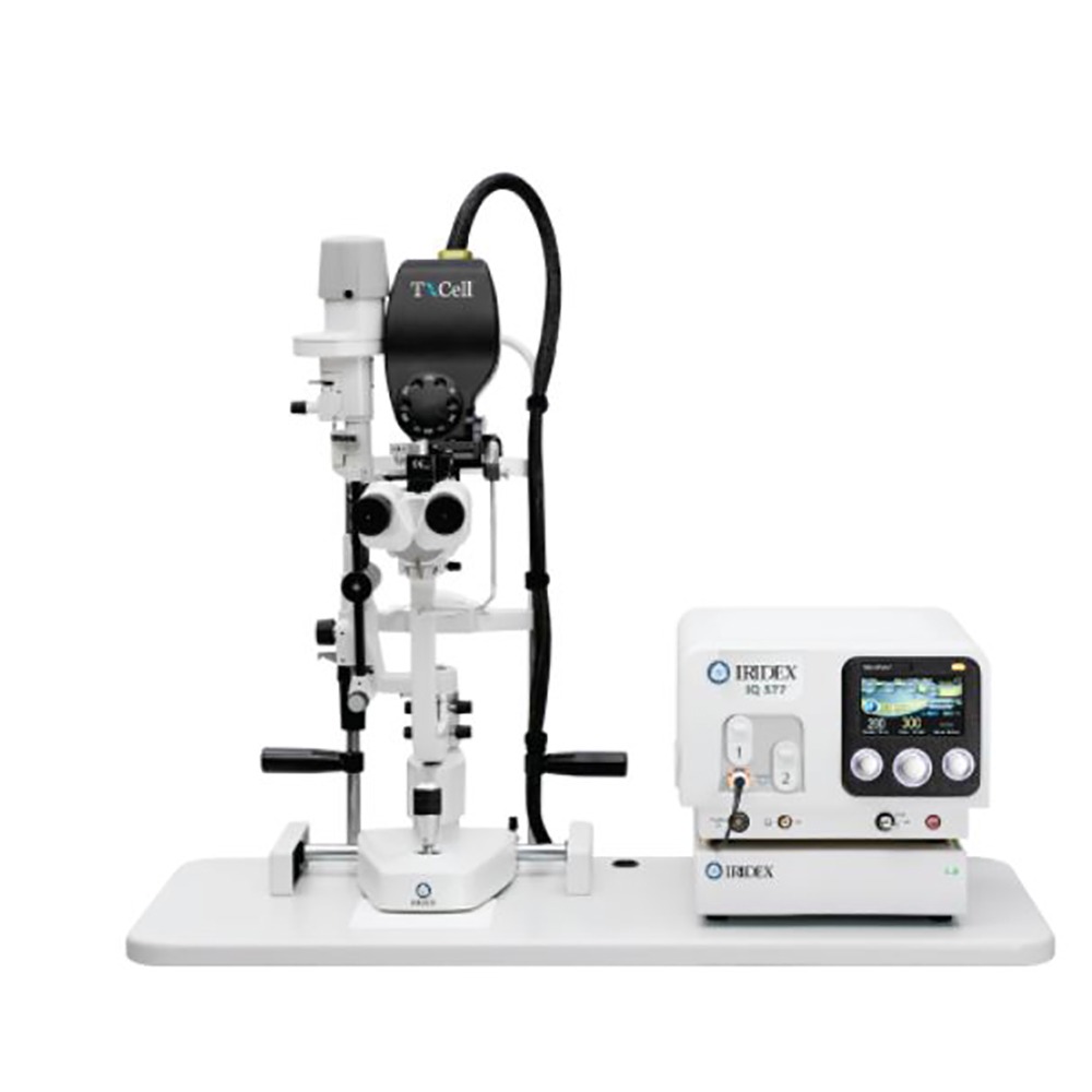
with the Iridex IQ 577. This is the top of the line latest laser equipment
for treatment of diabetic retinopathy. The Iridex IQ577 is a yellow light
laser.
This new laser option
requires less power and shorter pulse durations to treat the retinal bleeding.
This not only improves the comfort level but also can be used more frequently
to repair damage, than the older laser equipment.
The state-of-the-art Iridex
IQ577 is only used at Vision Express Surgery Centre at Karlione Court, Rodney
Bay, and this advanced yellow laser is not available anywhere else in Saint
Lucia.
“Vision Express is very oriented to
eye care, especially to patients with diabetes,” says Dr. Garcia. “It’s very
important because we have many people in Saint Lucia with diabetes. When a
person is diabetic for more than five years, they start to experience problems
with their retinas. It’s a very silent situation because until their central
vision is not affected, they do not realize that. That’s why it’s very
important for these patients to get routine eye checks.”
Vision Express also has a very
high-tech laser which can be used repeatedly. Previously, the machine
ophthalmologists used for treatment of diabetic
retinopathy left scars on the back of the eye. So once used, it could not
be used again. This is not the case with our new laser because we can use it
anytime.
Another unique treatment offered at
Vision Express is the eye injection. Some patients are unable to improve their
condition through laser treatment, so they need another method of treatment. As
such, the eye injection now available in Saint Lucia for the first time – only at
Vision Express.
“In the near future, we’ll be able to do retinal surgery for patients with diabetic retinopathy. It will be a huge plus for us and patients,” Dr. Garcia noted.
Dr. Garcia says the most common form
of diabetic retinopathy in Saint
Lucia is Type II Diabetes for which patients only need tablets most of the
time. However, if treated at a late stage, they will also need insulin. Most of
the time, our patients at Vision Express are middle-aged with a history of not
following proper dieting methods. Which is why he offers the following advice.
“My advice to anyone who has been diagnosed with diagnosed with diabetes is to get an eye examination,” Dr. Garcia said. “Quite often, they’ve been diabetic for a long time and didn’t realize it. Get tested early – the earlier the better. But at least once a year.”
At Vision Express, we offer patients
information about diabetes so that they are better informed. This means they
get a better picture of their condition. They also get the chance to get
treatment that was not available before in Saint Lucia.
Regular eye exams are very important in maintaining good eye health
and correcting any vision problems which may exist. People of all ages
should receive frequent checkups in order to prevent visual impairment
for conditions such as glaucoma, which can be managed if it is detected
in its early stages. In fact, according to a report from the World
Health Organization (WHO), there are about a billion people in the
world, living with vision problems which could have been prevented if
treatment was sought promptly.
Children for example should definitely get checked out to ensure that optimal eye health is maintained. As early as 6 months old, children should receive their first thorough eye screening by a professional eye specialist. Another screening should follow when the child turns 3 years of age and again at about 5 or 6 years old before joining first grade. Poor eyesight can adversely affect a child’s performance, causing headaches and an inability to focus on teachings and homework. A child’s hand eye coordination can also be affected and hinder the development of their fine motor skills and their ability to participate in sporting activities such as football, cricket and baseball.
Adults shouldn’t neglect their visits either, as they can also be affected by conditions such as presbyopia, which affects one’s ability to view close objects or read fine print on items such as newspapers and food labels. However, presbyopia is one such condition which can be easily managed by simply wearing glasses or contact lenses, which can be prescribed by an eye specialist after a routine checkup.
Seniors can also be
affected by poor vision and infrequent eye exams. As we grow older and
we age, our bodies go through changes which can develop into serious
health problems if we are not careful. Macular degeneration is one such
example of a visual impairment that adults over 60 years old experience,
when the macula becomes defective and images cannot be produced to be
sent to the brain. This can eventually lead to blurred vision and, in
the worst case scenario, individuals may become blind. This condition
may produce no symptoms during its early stages, which is why it is so
important that adults receive regular eye exams to protect their eye
health.
To order to maintain eye health, all adults, particularly
those from 18-60 years of age should receive eye exams every 2 years.
Individuals above 60 should be seen more frequently, at least once a
year, particularly due to conditions such as glaucoma and cataract which
are more common in older persons. Risk factors such as age, race,
diabetes and lifestyle choices such as smoking and lack of exercise can
increase one’s likelihood of developing eye diseases.
Frequent
checkups with the eye specialist should not be avoided. This painless
exam can not only help you to correct your vision, it can also detect
conditions in the early stages and prevent vision loss. A proactive
approach to your health will ensure that eye problems do not become a
regular part of your day and that can you live a life that you deserve.
Vision Express Medical , private surgery facility, is located in Karlione Court, Rodney Bay, St Lucia. Head Surgeon Dr Alejandro Garcia makes weekly consultation visits to all branches of Vision Express. To make it easier for patients in Castries and Vieux Fort to make full use of the diagnostic equipment and treatments available in the Rodney Bay Surgery Centre, Vision Express now offer free accompanied transport from Vieux Fort and Castries once a month.
Once a patient is diagnosed, for instance, with diabetic retinopathy, a full diagnosis and treatment plan can be made using the New Zeiss Cirrus OCT in Vision Express Medical, Rodney Bay. Follow on treatment, with laser to seal the blood vessels, damaged by diabetes, can be done using the new Iridex yellow laser equipment , or injections of Avastin or Lucentis can be given to help prevent further damage , both options also available in Vision Express Medical in Rodney Bay.
Glaucoma laser treatment is also sometimes an option, and the extent of damage for high intraoculat pressure can be checked with a full visual fields test or by using the OCT. Again both the visual fields equipment and the laser for glaucoma are available in Rodney Bay.
If you, or a loved one, are diagnosed with Cataracts, or decide to do Lens Replacement Surgery, the surgery facility at Rodney Bay offers full cataract removal with the Phaco equipment. On the surgery day it is preferable if a relative or friend accompanies the surgery patient, so this isn’t normally offered by Vision Express. But, prior to the surgery day a full check up is needed and measurement taken for the intraocular lens to be ordered. This is done using an ultrasound scan in Vision Express Medical in Karlione Court and the patient will be accompanied by their local Vision Express branch Manager.
None of the above diagnosis and treatments are painful, and being accompanied to do them with the local branch supervisor is the easiest way. As several patients will be traveling for treatment an early arrival in Rodney Bay and leaving mid afternoon is the preferred time frame. Patients who have had treatment are free to visit the Rodney Bay Malls and Vision Express Team will provide lunch and drinks throughout the day .
Vision Express work to provide the best service to all patients and are delighted to offer this new accompanied transport option to their customers in Vieux Fort and Castries to make full use of the range of the latest equipment for diagnosis and treatment of various eye problems under the excellent care of Retinal Surgeon , Dr Alejandro and his Professional Team.


 Diabetic retinopathy is caused by high blood sugar due to diabetes. Over time, having too much sugar in your blood can damage your retina, the part of the eye that detects light and sends signals to the brain through a nerve in the back of the eye (optic nerve).