The 411 On Retinal Detachment

Eye Diagram - VE St Lucia

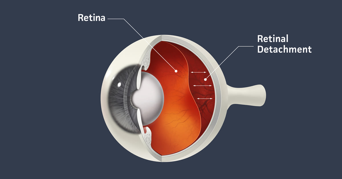
The retina is the light-sensitive tissue lining the back of the inside of the eye. It’s made up of different layers, including the light-sensing cells known as photoreceptors that are essential for sight. A layer of underlying supporting tissue contains blood vessels that supply the photoreceptors with the food and oxygen they need to stay healthy.

The retina functions as the line of communication between our eyes and the world around us by sending visual messages through the optic nerve to the brain. A healthy, intact retina is crucial for clear vision. But if it becomes detached, you can permanently lose your eyesight.

Types of Retinal Detachment

There are three ways a retina can detach:

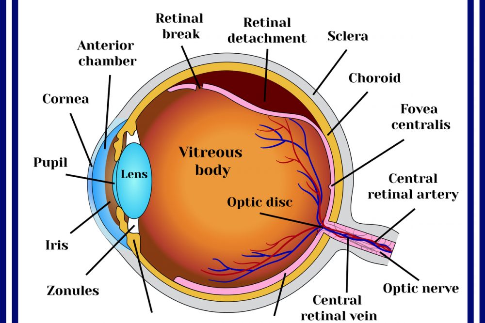
  • Rhegmatogenous – A tear allows fluid to get under the retina, separating it from the retinal pigment epithelium (RPE), which nourishes the retina. This is the most common form of retinal detachment.
  • Tractional – Scar tissue on the retina’s surface contracts, causing it to separate from the RPE.
  • Exudative – Fluid leaks into the area underneath the retina. This type is usually caused by inflammatory diseases or eye injuries.

Can high blood pressure cause retinal detachment?

Along with causing heart and kidney problems, untreated high blood pressure can also affect your eyesight and lead to eye disease. Hypertension can cause damage to the blood vessels in the retina. This eye disease is known as hypertensive retinopathy.

What are the risk factors of a Detached Retina?

While retinal detachment is more common in people older than 40, especially men, it can occur at any age. Other risk factors include:

  • Family history
  • Having an eye disease or injury
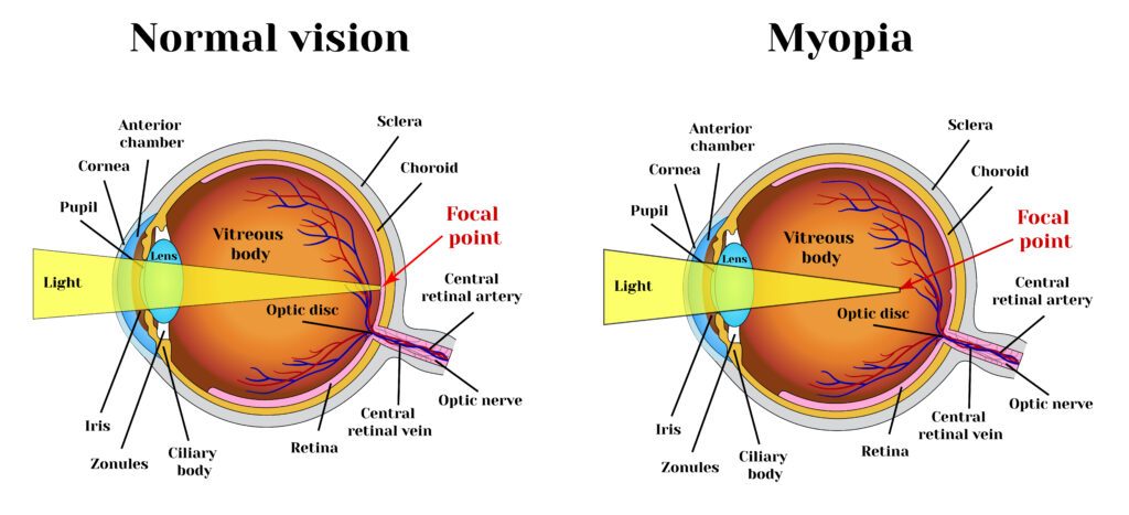
  • Nearsightedness
  • Previous eye surgery
  • Weak areas in the retina

Symptoms of Retinal Detachment

Retinal detachment is painless; however, various warning signs point to its existence, including:

  • Blurred vision
  • Flashes of light in one or both eyes
  • Gradually reduced peripheral vision
  • Shadow over visual field
  • Sudden appearance of floaters

It is advised that should you experience any of these symptoms, you should seek medical attention immediately. At Vision Express, our ophthalmologist may diagnose retinal detachment via either of the following procedures:

  • Retinal Examination – In this procedure, a tool with a bright light and a special lens (ophthalmoscope) is used to examine the back of your eye, thus allowing your doctor to see retinal detachments, holes or tears.
  • Ultrasound Imaging – This procedure is used if bleeding has occurred in your eye making it difficult to see the retina.

Should your doctor fail to see a detachment, he or she may ask you to return in a few weeks to confirm that your eye has not developed any tears. Should you continue to experience symptoms, seek medical treatment immediately.

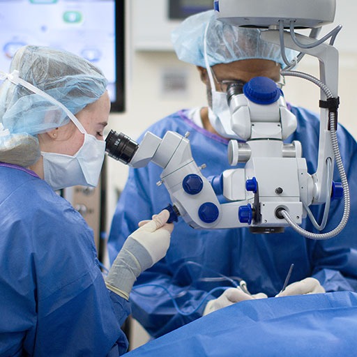
How is a Detached Retina treated?

Treatment for a detached or torn retina requires surgery. Tears are treated by sealing the retina to the back wall of the eye using these surgical treatments:

  • Freezing treatment (cryopexy): In this procedure, your eye surgeon uses a probe to freeze the retina around the tear. The scar helps secure the retina to the eye wall.
  • Laser surgery (photocoagulation): In this procedure, your ophthalmologist uses a laser to make small burns around the tear, creating scars. This seals the retina to the underlying tissue and helps prevent future detachment.

If your retina has detached, you will also need to have surgery to reattach it in its proper position. Otherwise, you could go blind. There are three surgical methods:

  • Pneumatic Retinopexy – In this procedure, a gas bubble is injected into the eye, pushing the tear back against the eye wall. Subsequently, your ophthalmologist will ask you to maintain a certain head position for several days.
  • Scleral buckle – In this procedure, a flexible band is placed around the eye to counteract the force pulling the retina out of place. Your doctor will drain the fluid underneath, allowing the retina to settle back into its normal position.
  • Vitrectomy – In this procedure, the vitreous gel is removed from the eye to prevent pulling. It is replaced with a gas or oil bubble to keep the retina in place until your body’s own fluids can fill the space. A gas bubble will go away on its own while an oil bubble must be surgically removed.

Following surgery, your vision may take months to improve and may not ever fully return. In some cases, the retina cannot be reattached and the eye will continue to lose sight.

How quickly should a Detached Retina be treated?

If your retina has detached, you will need surgery to repair it preferably within days of a diagnosis. The type of surgery your surgeon recommends will depend on several factors, including how severe the detachment is. 

If you need to know more about retinal detachment, contact Vision Express today to make an appointment.

What is Myopia?

Myopia - Vision Express St Lucia

Myopia, also referred to as nearsightedness, is a vision condition whereby close objects can be seen clearly while distant objects appear blurred. As such, people with myopia have difficulty seeing a movie or the television screen clearly, a blackboard in class or while driving in traffic.

Myopia is caused when the eyeball is too long or the cornea (the clear front cover of the eye) is too curved, resulting in the light entering the eye to not focusing correctly and distant objects appearing blurred.

While the exact cause of myopia remains unknown, significant evidence suggests that many people inherit myopia or the tendency to develop the condition. There is an increased chance that children will be nearsighted if one or both parents are nearsighted.


While the tendency to develop myopia may be inherited, its actual development may be affected by how a person uses his or her eyes. People who spend considerable time reading, working at a computer, or doing other intense close visual work are more likely to develop myopia.

Myopia first occurs in school-age children. Since the eye continues to grow during childhood, it typically progresses until about age 20. However, myopia may also develop in adults due to visual stress or conditions such as diabetes.


Myopia can also be caused by environmental factors or other health problems:

  • Some people may experience blurred distance vision only at night. With “night myopia”, low light makes it difficult for the eyes to focus properly. Also, the increased pupil size during dark conditions allows more peripheral, unfocused light rays to enter the eye.
  • People who do excessive amounts of near-vision work may experience a false or “pseudo” myopia. Their blurred distance vision is caused by overuse of the eyes’ focusing mechanism. After long periods of near work, the eyes are unable to refocus to see clearly in the distance. Clear distance vision usually returns after resting the eyes. However, over time, constant visual stress may lead to permanent reduction in distance vision.
  • Symptoms of myopia may also be a sign of variations in blood sugar levels in people with diabetes or may be an early indication of a developing cataract.

 At Vision Express, an optometrist can determine the cause of the vision     problems through a comprehensive eye exam.

Diagnosis for myopia 

Testing for myopia may use several procedures to measure how the eyes focus light and to determine the power of any optical lenses needed to correct the reduced vision.

As part of the testing, you will identify letters on a distance chart to measure visual acuity, written as a fraction, such as 20/40. The top number of the fraction is the standard distance at which testing is performed. The bottom number is the smallest letter size read. Someone with 20/40 visual acuity would have to get within 20 feet to identify a letter that could be seen clearly at 40 feet in a “normal” eye. Normal distance visual acuity is 20/20, although many people have 20/15 (better) vision.

Using an instrument called a phoropter, the optometrist places a series of lenses in front of your eyes and measures how they focus light using a handheld lighted instrument called a retinoscope. The doctor may also use an automated instrument that evaluates the focusing power of the eye. The power is then refined based on your responses to determine the lenses that allow the clearest vision.

Your doctor can conduct this testing without using eye drops to determine how the eyes respond under normal seeing conditions. In some cases, such as for patients who can’t respond verbally or when some of the eye’s focusing power may be hidden, the doctor may use eye drops which temporarily keep the eyes from changing focus during testing.

Using the information from these tests, alongside the results of other tests of eye focusing and eye teaming, your doctor can determine if you have myopia and also the power of any lens correction needed to provide clear vision. Once testing is complete, your doctor can discuss treatment options.


Treatment for myopia

People with myopia have several options available to regain clear distance vision, including:

Eyeglasses: Eyeglasses are the primary choice for correction for most people with myopia. Depending on the amount of myopia, you may only need to wear glasses for certain activities such as watching a movie or driving a car. If you are very nearsighted, you may need to wear glasses all the time.

Generally, a single-vision lens is prescribed to provide clear vision at all distances. However, patients over age 40 or children and adults whose myopia is due to the stress of near-vision work may need a bifocal or progressive addition lens. These multifocal lenses provide different powers or strengths throughout the lens to allow for clear vision in the distance and up close.

Contact lenses: For some individuals, contact lenses offer clearer vision and a wider field of view than eyeglasses. However, since contact lenses are worn directly on the eyes, they require proper care to safeguard eye health.

Ortho-k or CRT: Another option for treating myopia is orthokeratology (ortho-k), also known as corneal refractive therapy (CRT). In this non-surgical procedure, you wear a series of specially-designed rigid contact lenses to gradually reshape the curvature of your cornea, the front outer surface of the eye. 

The lenses place pressure on the cornea to flatten it and this changes how light entering the eye is focused. You wear the contact lenses for limited periods, such as overnight, before removing them. People with mild myopia may be able to temporarily obtain clear vision for most of their daily activities.

Laser procedures: Laser procedures such as LASIK (laser in situ keratomileusis) or PRK (photorefractive keratectomy) are also possible treatment options for myopia in adults. A laser beam of light reshapes the cornea by removing a small amount of eye tissue. The amount of myopia that PRK or LASIK can correct is limited by the amount of corneal tissue that can be safely removed.

In PRK, a laser removes a thin layer of tissue from the surface of the cornea in order to change its shape and refocus light entering the eye.

LASIK removes tissue from the inner layers, but not from the surface, of the cornea. To do this, a section of the outer corneal surface is lifted and folded back to expose the inner tissue. A laser then removes the precise amount of corneal tissue needed to reshape the eye. Then, the flap of outer tissue is placed back in position to heal.

Other refractive surgery procedures: People who are highly nearsighted or whose corneas are too thin for laser procedures may be able to have their myopia surgically corrected. A doctor may be able to implant small lenses with the desired optical correction in their eyes. The implant can be placed just in front of the natural lens (phakic intraocular lens implant), or the implant can replace the natural lens (clear lens extraction with intraocular lens implantation). This clear lens extraction procedure is similar to cataract surgery but occurs before a cataract is present.

Vision therapy for people with stress-related myopia: Vision therapy is an option for people whose blurred distance vision is caused by a spasm of the muscles that control eye focusing. Various eye exercises can improve poor eye focusing ability and regain clear distance vision.

If you have myopia, you have a variety of options to correct your vision problem. In consultation with your doctor at Vision Express, you can select the treatment that best meets your visual and lifestyle needs.